

 Online Doctor Appointment
  Health Check up Package
 Contact us
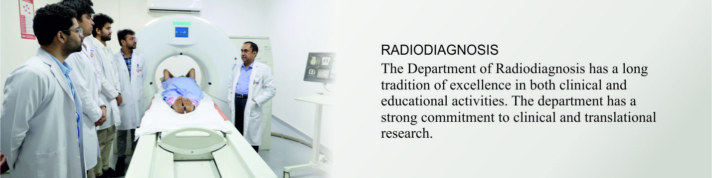
 DEPARTMENT OF RADIODIAGNOSIS
The Department of Radiodiagnosis has a long tradition of excellence in both diagnostic and educational activities. We are guided by the mission to maintain excellence in education, research and time and can find illness early – which can be treated most effectively. The department has state-of-the-art advanced equipments – two CT scans, 128 slice dual source and 32 slice spiral CT scans for all advanced imaging and angiography studies including cardiac angiography, 3 Tesla wide bore MRI and one 1.5 T (Vario, 18 Channel, Siemens). The department has experienced and dynamic team of radiologists and technician providing diagnostic and interventional support round the clock.
FACULTY
Carousel contents not found!RESEARCH & PUBLICATION
Madhok R, Aggarwal AK, Aggarwal A. Epidemiological study of epilepsy in northern india through evaluation by 3 tesla MRI. Int J Res Health Sci. 2015; 3(2): 256-66.
Madhok R, Rastogi S. Role of 3 tesla magnetic resonance cholangiopancreatography in obstructive jaundice with cyto/ histopathological or surgical correlation. Int J Sci Stud. 2015; 3(2): 1-7.
Madhok R, Rastogi SK, Aggarwal A, Kapoor A, Rastogi S. Determining The Indications & Role Of Screening/ Diagnostic Mammography In Breast Cancer In Women below 40 yrs & Contribution of Ultrasound As An Adjunct To Mammography: A Retrospective Study. Int J Sci Stud. 2015; 2(11): 1-7.
Madhok R, Aggarwal A. Comparison of 128- Slice Dual Source CT Coronary Angiography with Invasive Coronary Angiography. J Clin Diagn Res. 2014; 8(6): 8-11.
Tapasvi C, Prajapati N, Madhok R, Gupta AK, Taneja V, Aggarwal A. Evaluation of Bowel wall Thickening by Computed Tomography to differenties. J Clin Diagn Res. 2014; 8(11): 9-12.
Gupta A, Madhok R, Aggarwal A. Role of magnetic resonance imaging in neoplastic diseases of spine. Int J Res Health Sci. 2015; 3(I); 5-10.
Das G, Gupta AK, Aggarwal A. Assessment of lower limb arteries by Doppler sonography in diabetic patients. Int J Res Health Sci. 2015; 3(I); 18-23.
Prajapati N, Madhok R, Tapasvi C, Prashad U, Rastogi S. High frequency and color Doppler ultrasound evaluation of scrotal and testicular pathologies in indexed journal. Int J Res Health Sci. 2(1): 153-61.
Prajapati N, Madhok R, Rastogi S, Rawal D, Tapasvi C. 3 Tesla MRI Evaluation of febrile and unconscious patients with CSF analysis correlation in an endemic zone for herpes and Japanese encephalitis. Ind J Basic App Med Res. 2014; 3(4): 8-14.
Prajapati N, Rastogi SK, Tapasvi C, Taneja V. Scrotal and testicular masses evaluation by high resolution Ultrasonography. Int J Res Health Sci. 2014; 4(1):430-8.
Pant HC, Prajapati N, Jain A, Taneja V. Ultrasound and MRI evaluation of hepatobiliary tumors with Histopathological correlation. Int J Res Health Sci. 2015; 4(3):324-32.
12 Madhok R, Taneja V. Role of sonosalpingogram in correlation to hysterosalpingogram in assessment of infertility. Int J Reprod Contracept Obstet Gynaecol. (Accepted for June 2016).