
Single Photon Emission
Computed Tomography
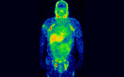
Similar to PET, single photon emission computed tomography (SPECT) uses radioactive tracers and a scanner to record data that a computer constructs into two- or three-dimensional images. The tracer is what allows doctors to see how blood flows to tissues and organs & provide them with functional information of that particular organ which can be read together with other anatomical imaging information to manage the treatment of the patients in a better way.
A small amount of a radioactive drug is injected into a vein and a scanner is used to make detailed images of areas inside the body where the radioactive material is taken up by the cells. SPECT can give information about blood flow to tissues and chemical reactions (metabolism) in the body.
What Does a Spect Scan Show?
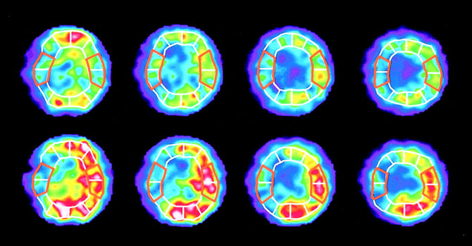
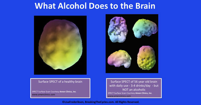
A SPECT scan is primarily used to view how blood flows through arteries and veins in the brain. Tests have shown that it might be more sensitive to brain injury than either MRI or CT scanning because it can detect reduced blood flow to injured sites
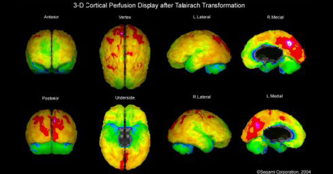
SPECT scanning is also useful for presurgical evaluation of medically uncontrolled seizures The test can be performed between seizures (interictal) or during a seizure (ictal) to determine blood flow to areas where the seizures originate.
SPECT scanning is also useful for presurgical evaluation of medically uncontrolled seizures The test can be performed between seizures (interictal) or during a seizure (ictal) to determine blood flow to areas where the seizures originate.
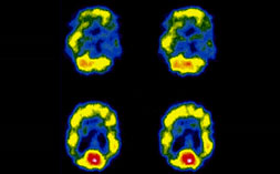
A SPECT scan of a patient with uncontrolled complex partial seizures. The temporal lobe on the left side of the brain shows less blood flow than the right, confirming for the surgeon the nonfunctioning area of the brain causing seizures
It is used for assessing blood flow to the brain helping in documenting early dementia changes; for identifying seizure focus in intractable epilepsy cases using ictal-SPECT, and for suspected tumor recurrences. Can also be used in few neuropsychiatric disorders such as schizophrenia, depression etc.
Differential Diagnosis of Thyrotoxioicosis Graves Disease Sub acute Thyroiditis, Auto nornous functioning thyroid nodule (AFTN), Thyrotoxicosisfactitia Determination of functional status of thyroid nodule Cold Nodule (Avscular /Hypervascular) Hot Nodule Detection of ectopic thyroid tissue (ex. Lingual Thyroid) Evaluation of hypothyroldism in new born.
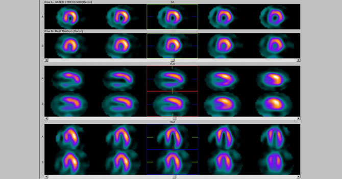
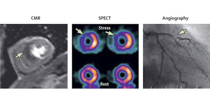
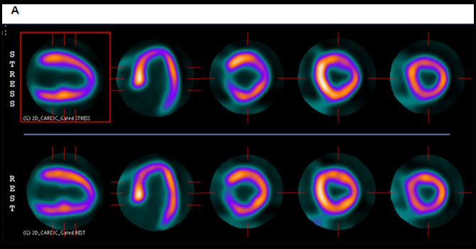
Myocardial Perfusion Imaging/Stress Thallium
Evaluation of chronic angina.Confirmation of coronary stenosis detected on cath. angio (Flame test of Gold standard). Evaluation of patients before CABG (by-pass) or Angioplasty to assess the myocardial perfusion and functional aspect of heart. Follow up of patients post CABG (by pass) or Angioplasty, Routine heart check up
Yocardial Viability Test
To check for the myocardial viability before any planned intervention. To assess the prognosis of the planned intervention. As an alternative to Stress thallium for patients who can’t undergo stress.
Muga/Radionuclide Angiography
Calcution of LVEF (especially in patients with poor Echo window) Evaluation during cardiatoxic chemotherapy and follow up afterwards. Post stem cell treatment.
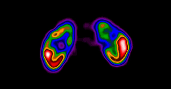
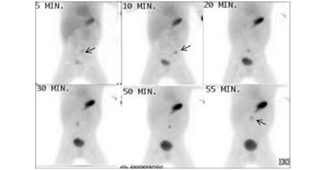
Renal Dynamic Scan (dtpa)
Functional assessment (GFR evaluation) for ex. Pre chemotherapy evaluation, Donor kidney evaluation (before transplant), Renal failure assessment, Transplanted Kidney evaluation, Ureteral obstruction/PUJ obstruction / Hydronephrosis
Captopril Renogram
To rule out renal artery stenosi (RAS)
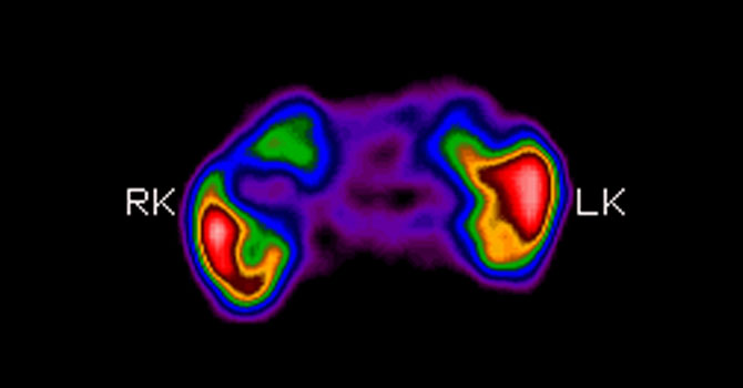
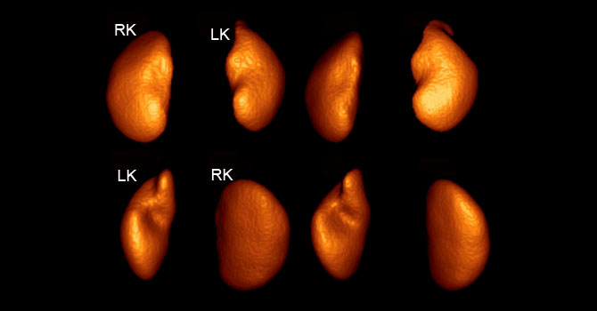
Renal Dmsa Scan
Missing Kidney, Scarring, Pyelonephiritis
DRCG Scan
Missing Kidney, Scarring, Phyelonephritis
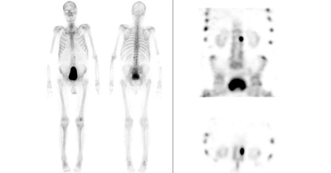
Single Phase Bone Scan
Metastatic bone tumours, Benign and malignant primary bone tumors, Metabolic bone disease, Paget’s disease
Triple Phase Bone Scan
Avascular necrosis of bone, Osteoarthiritis (to differentiate between inflammatory & non inflammatory arthritis), Reflex sympathetic dystrophy syndrome (RSD)/ Chronic regional pain syndrome (CRPS), Tubercular Osteomylitis, Stress fracture, Sport injury of bone, Prosthesis loosening/ infection post THR/TKR.