Single Photon Emission
Computed Tomography
Similar to PET, single photon emission computed tomography (SPECT) uses radioactive tracers and a scanner to record data that a computer constructs into two- or three-dimensional images. The tracer is what allows doctors to see how blood flows to tissues and organs & provide them with functional information of that particular organ which can be read together with other anatomical imaging information to manage the treatment of the patients in a better way.
To detect and locate hyper functioning parathyroid adenomas.
Preparation:
To stop ant thyroid drugs at least 72 hrs before scan.
Book nowTo assess Thyroid function and nature of thyroid nodules.
Book nowDTPA Renal Scan (Renal Dynamic Scan) test helps in evaluating renal function, blood flow and also determines obstructive pathologies of entire urinary system. Helps evaluation of transplanted kidneys as well
Preparation:
Adequate hydration required except when on fluid restriction.
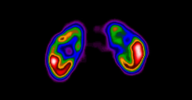
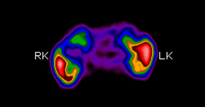
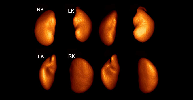
Book nowThis scan helps to assess renal morphology, structure (scar) and function. No age restriction.
Preparation:
Adequate hydration required except when on fluid restriction.
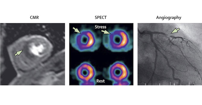
Book nowDone to assess ischemia in various territories of left ventricle of heart.
Preparation:
Selective withdrawal of beta-blockers, CCBs, caffeinated drinks, Nitrates for 24-48 hrs
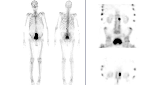
Book nowDone to assess both metabolic and metastatic pathologies of entire skeletal system.
Preparation:
Adequate hydration
Book nowDone to assess left ventricle Ejection Fraction of heart in patients on chemotherapy.
Book nowFunctionally detects ectopic spleen or residual spleen post Splenectomy.
Book nowHIDA scan is a Gamma Camera imaging procedure used to diagnose varied maladies of the liver, gallbladder and bile ducts.
Preparation:
4hrs fasting required
Book nowImaging technique to identify the lymphatic obstruction. Also helps identify sentinel lymph node in various malignancies.
Preparation:
4 hrs fasting required
Book nowPerformed to identify urinary reflux.
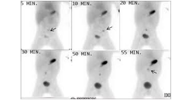
Book nowHelps assess location of gastrointestinal blood loss with high sensitivity.
Preparation:
Overnight fasting
Book nowHelps identify and locate misplaced gastric mucosa.
Preparation:
On Antacid for 48 hrs before scan
Book nowAccesses movement along GI system in most physiologic conditions.
Preparation:
Overnight fasting. Pro-kinetic withheld.
Book now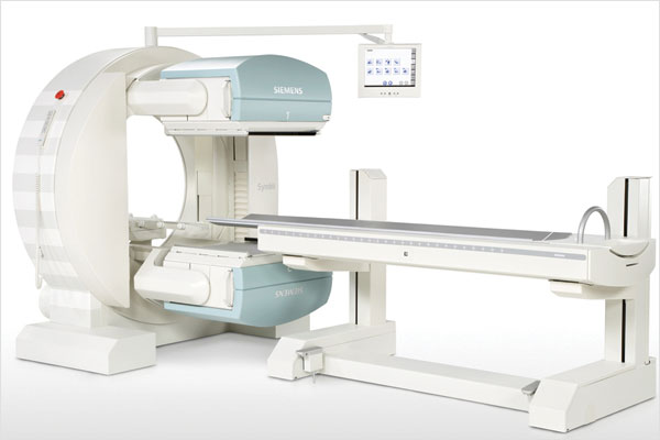
A small amount of a radioactive drug is injected into a vein and a scanner is used to make detailed images of areas inside the body where the radioactive material is taken up by the cells. SPECT can give information about blood flow to tissues and chemical reactions (metabolism) in the body.
What does a SPECT scan show?
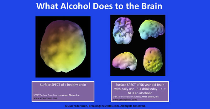
A SPECT scan is primarily used to view how blood flows through arteries and veins in the brain. Tests have shown that it might be more sensitive to brain injury than either MRI or CT scanning because it can detect reduced blood flow to injured sites
SPECT scanning is also useful for presurgical evaluation of medically uncontrolled seizures The test can be performed between seizures (interictal) or during a seizure (ictal) to determine blood flow to areas where the seizures originate.
 SPECT scanning is also useful for presurgical evaluation of medically uncontrolled seizures The test can be performed between seizures (interictal) or during a seizure (ictal) to determine blood flow to areas where the seizures originate.
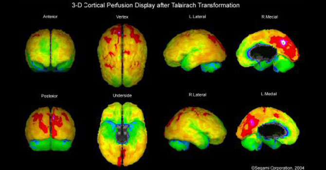
 A SPECT scan of a patient with uncontrolled complex partial seizures. The temporal lobe on the left side of the brain shows less blood flow than the right, confirming for the surgeon the nonfunctioning area of the brain causing seizures