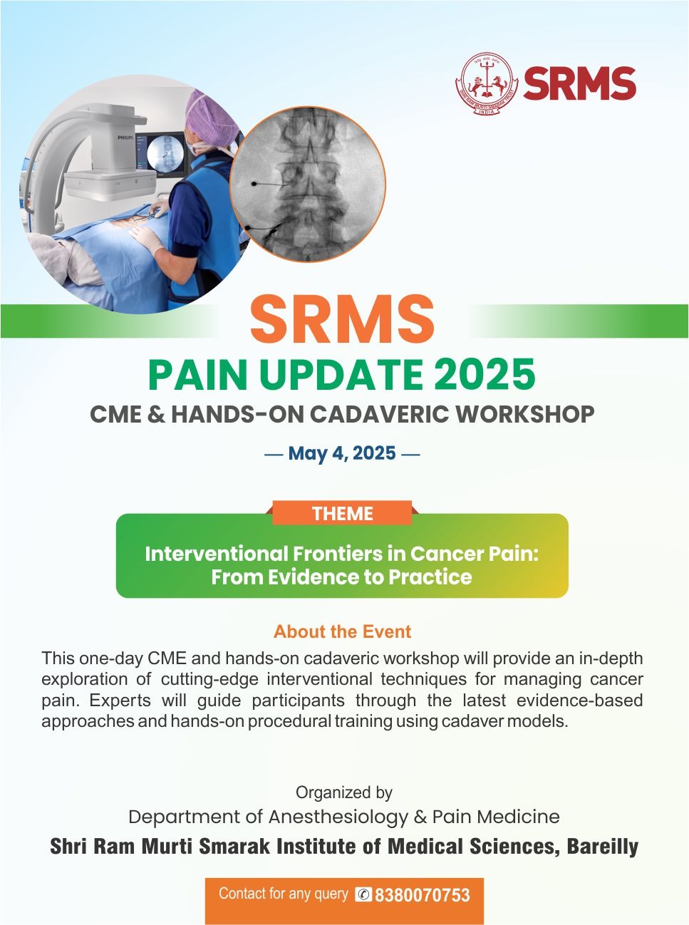
Flow Motion, the End of Stop and Go the Most Advanced

PET/CT in India

This is the era of Fusion imaging i.e fusing the functional information with the anatomical images. The functional information gives us the metabolic status of the particular organ whereas anatomical images provides us with the structural/morphological changes

Here at SRMS FIMC, we have got one of the most advanced fusion imaging modality in the form of Biograph mCT Flow PET-CT scanner which for the first time in this part of world, can provide us the most detailed information about any functional abnormality in the human body; basically in oncology but not limited to; with some diseases of neurology and cardiac origin as well.


Using Flow Motion Technology

Overcome Today’s PET/CT Challenges

Until now, PET examinations have been performed in sequential bed positions, alternating between acquisition and patient table motion. As such, image quality and quantitative accuracy have always been restricted, and scans often resulted in higher dose and greater patient anxiety. Powered by Siemens’ revolutionary FlowMotion technology, Biograph mCT Flow is the world’s first PET/CT system to eliminate the demand for stop-and-go imaging.

With Flow Motion examinations, parameters such as speed, image resolution and motion management can be easily adjusted to the precise dimensions of organs and routinely


FlowMotion technology eliminates overlapping bed acquisitions while maintaining uniform noise sensitivity across the entire scan range, enabling


FlowMotion allows easy anatomy-based scan planning where only the desired area is scanned.


Biograph mCT Flow is the first-and-only PET•CT to combine a large 78 cm bore with Flow Motion’s
continuous sense of scan progress, offering the


Unique Features

Finest Volumetric Resolution

Biographm mCT Flow with its OptisoHD detection system optimizes each component of the imaging chain.In addition to that, this comes with single source dual-energy CT capabilities for improved lesion contrast.

Virtually Freeze Motion

Through an innovative combination of hardware and software, HDoChest virtually freezes respiratory motion, enabling full HD lesion detection and accurate standard uptake value (SUV) quantification.The five-year survival rate for NSCLC may more than double if caught earlier, benefiting up to 85% of lung cancer patients. 3FlowMotion simplifies integration of HDoChest into every scan.

Precise Organ Imaging

Examination parameters such as speed, image resolution and motion management can be easily adjusted to the precise dimensions of organs.

Hi-Rez Increases Accuracy

Improved accuracy can change patient management in up to 40% of head and neck cancer cases.4 FlowMotion technology additionally provides simple incorporation of Hi-Rez into the PET scan protocols. This PET reconstruction with the industry’s highest1400x400 matrix can increase resolution potentially resulting in improved detection.

Oncological Applications

  • Staging and prognostication of various malignancies.
  • Suspected relapse where MR is equivocal to inform decisions regarding surgery or radiotherapy planning.
  • Differentiation between radiation necrosis and tumor recurrence.
  • Assessment of patients with elevated thyroglobulin levels and negative iodine scintigraphy, with suspected recurrent disease in case of Thyroid cancer.
  • Characterisation of a solid solitary pulmonary nodule (SPN).
  • To guide biopsy site in patients with large nodal/ tumoral mass where chances of necrosis are high.
  • Prior to bone marrow transplant, in cases of lymphoma; to assess volume of disease and suitability for transplant
  • To determine extent and identify a suitable biopsy site in patients with low-grade lymphomas in whom there is suspected high-grade transformation.
  • To detect an occult primary tumour in selected patients with non-metastatic manifestations of neoplastic disease (Para Neoplastic Syndrome) when other imaging is negative or equivocal
  • Detection of the primary site when imaging and histopathology has failed to show a primary site (carcinoma of unknown primary, CUPS), where the site of tumour will influence choice of chemotherapy.

Non-Oncological Applications

Neurology
  • Pre-surgical assessment of medically refractory seizures where MR is normal, equivocal or conflicts with EEG localization.
  • Evaluation and differentiation of various types of dementia.

Cardiology

Assessment of myocardial viability in patients with Ischaemic Heart Disease and poor left ventricular function being considered for revascularization in combination with perfusion imaging with sestamibi/tetrofosmin/ thallium.

Infection Imaging
  • Detection of site of focal infection in immuno-compromised patients or problematic cases of infection.
  • Evaluation of vascular graft infection in selected cases provided sufficient time has elapsed since surgery.
Sarcoidosis

Assessment of activity and distribution of disease at baseline in highly selected cases where there is diagnostic uncertainty using conventional imaging (e.g. suspected cardiac sarcoidosis). Assessment of disease response where other measures to monitor response are unhelpful and/or in patients with disease resistant to treatment.

Vasculitis

Evaluation of suspected vasculitis in selected cases; for example, to determine the extent & distribution of the disease activity.

Pyrexia of unknown origin (PUO)

To identify the cause of a PUO where conventional investigations have not revealed a source.

Image Gallery

  • PET-CT SCAN
  • PET-CT SCAN
  • PET-CT SCAN
  • PET-CT SCAN
  • PET-CT SCAN
  • PET-CT SCAN
  • PET-CT SCAN
  • PET-CT SCAN
  • PET-CT SCAN
  • PET-CT SCAN
  • PET-CT SCAN