Explore SRMSIMS
Medical News & Events
Upcoming Events

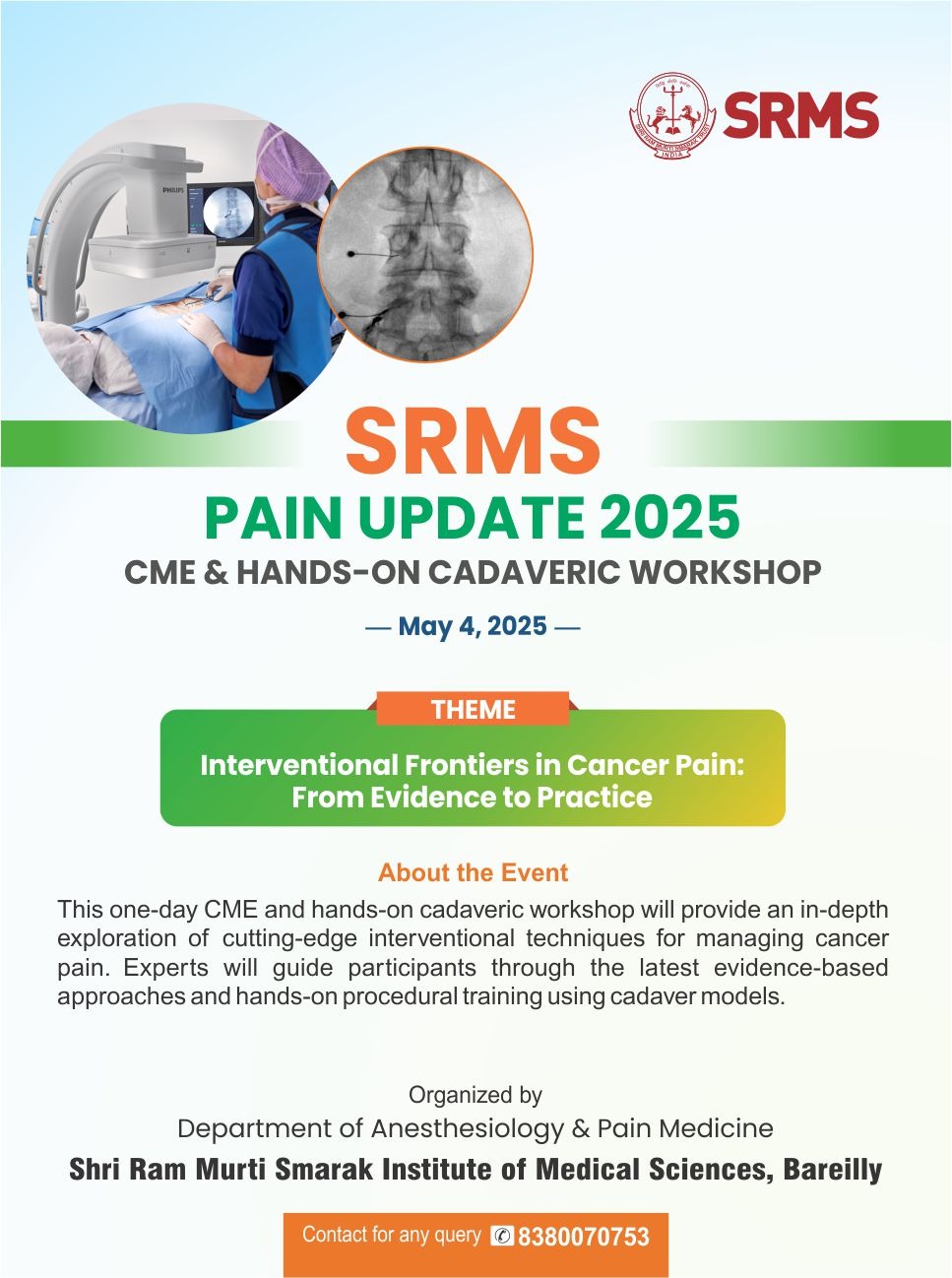
SRMS
PAIN UPDATE 2025
CME & HANDS-ON CADAVERIC WORKSHOP
Interventional Frontiers in Cancer Pain : From Evidence to Practice
Information Under NMC
Medical News & Events
Upcoming Events

SRMS
PAIN UPDATE 2025
CME & HANDS-ON CADAVERIC WORKSHOP
Interventional Frontiers in Cancer Pain : From Evidence to Practice
Quick Links
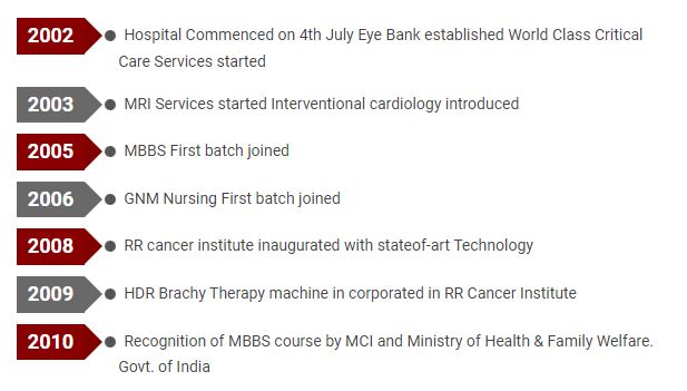
Milestones of SRMSIMS

LEADING IN PRECISION HEALTH

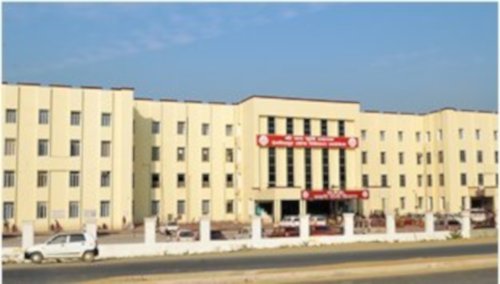
SRMSIMS Hospital
Shri Ram Murti Smarak Institute of Medical Sciences is one of the best Hospital in Bareilly which has most modern 950 bed, centrally air-conditioned, Multi Super Speciality, Tertiary Care and a Trauma Hospital established on 4th July’ 2002. The Hospital has 600 teaching beds for teaching and training of students pursuing their MBBS Programme. The treatment of the patients on these beds is done free of cost. The Hospital is well equipped with facilities to provide quality teaching & training to the students in various departments. Students during their clinical training are exposed to variety of patients so as to make them competents in the field of medicine. During their training students are also exposed to behavioural & managerial skills required in todays world for becoming a complete medical practitioner.


